Fraunhofer Institute for Photonic Microsystems
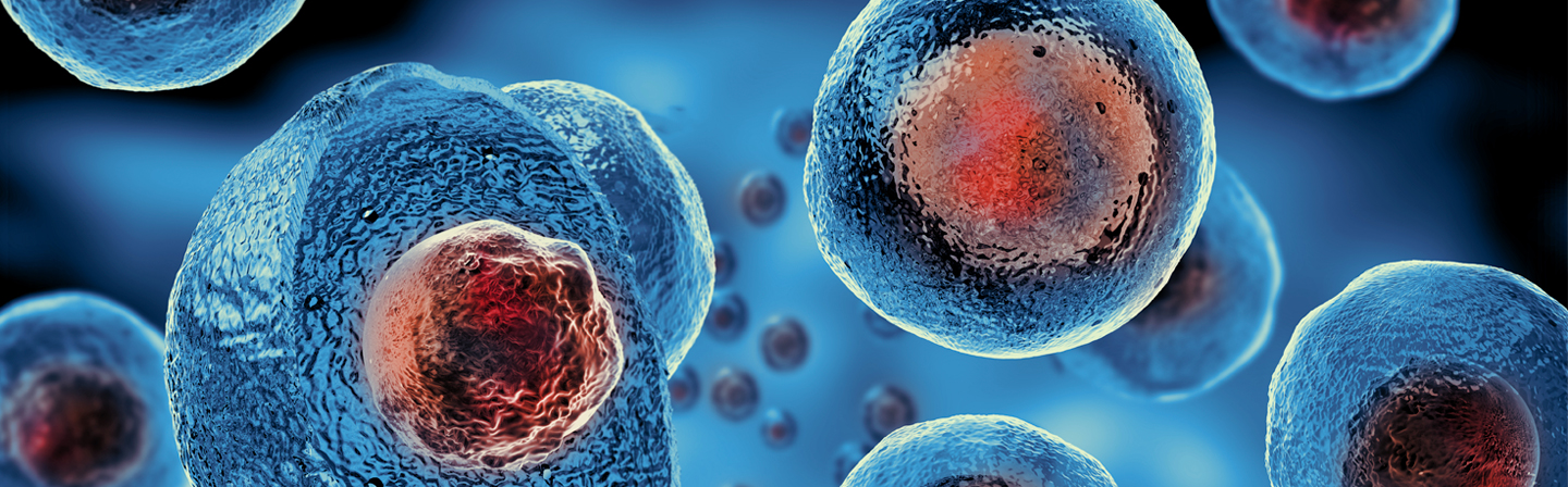
Confocal Microscope for Rapid Detection of Tumor Boundaries during Surgery


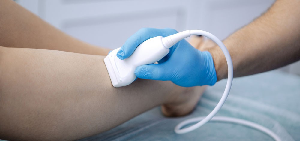
Each year, around 500,000 people in Germany are diagnosed with cancer. While diagnostic tools and treatment methods are continually improving, surgeons still face a major challenge: determining in real time whether a tumor has been completely removed during surgery. Currently, it is standard practice to extract tissue samples from the resection margin and send them to pathology for analysis - a process that typically takes up to 20 minutes.
What’s needed is a fast, reliable, and on-site solution that can be used directly in the operating room. To meet this demand, researchers from Fraunhofer IPMS and Fraunhofer IZI have developed a new confocal laser scanning microscope for intraoperative tumor diagnostics.
Differentiating Tumor from Healthy Tissue in Real Time
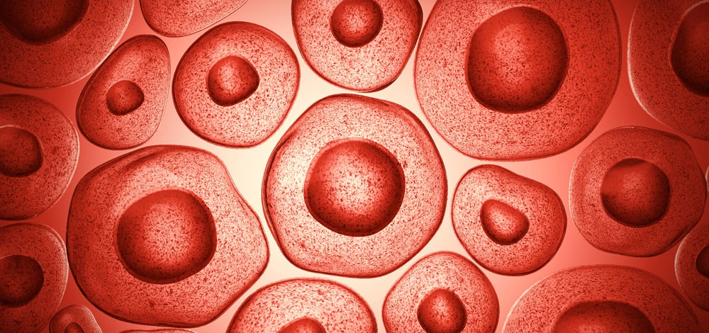
A key challenge in tumor surgery is clearly distinguishing between healthy and cancerous tissue. The goal is to achieve tumor-free resection margins while preserving as much healthy tissue as possible - a critical requirement especially in sensitive areas such as the brain.
Current intraoperative diagnostic methods such as histology are limited: they take time, may not work on certain tissue types like bone, and typically do not provide 3D visualization of tumor margins. This has created a strong need for a rapid optical method that can reliably differentiate tissue types during surgery.
MEMS-Based Confocal Microscopy with Fluorescent Tumor Markers
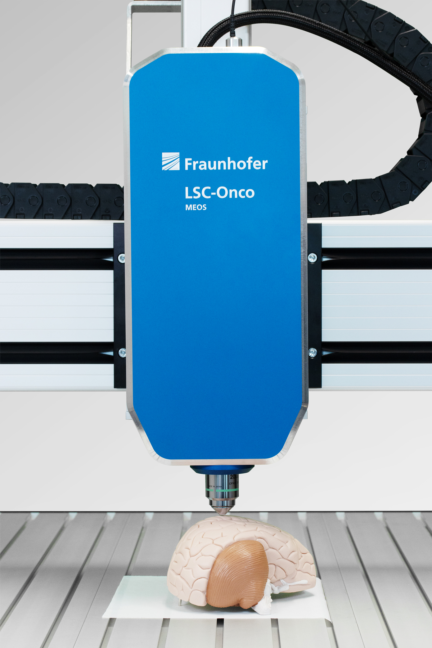
To address this need, Fraunhofer IPMS and Fraunhofer IZI have developed a MEMS-based confocal laser scanning microscope combined with a fluorescent antibody staining technique for tumor cells. The system is designed to identify tumor boundaries with high spatial precision, aiding surgeons in preserving critical structures like brain tissue and arteries.
The process begins by staining the tumor tissue with fluorescence-labeled antibodies, developed by Fraunhofer IZI. Then, the microscope, which is equipped with a MEMS scanning mirror from Fraunhofer IPMS, captures real-time images of the tissue surface. The mirror deflects light in both x and y directions to produce live, high-resolution images.
Key technical specifications:
- Lateral resolution: < 1.0 μm
- Field of view: 200 x 200 μm² (960 x 960 pixels)
- Z-axis imaging: 2000 μm range, 5 nm step size
First Demonstrator and Next Steps
In 2021, a functional demonstrator of the microscope was successfully tested at the Fraunhofer Center MEOS in Erfurt, using tissue samples provided by clinical partner Helios Klinikum Erfurt.
Looking ahead, future developments will include:
- AI-assisted detection of tumor margins
- Robotic integration to support surgical workflows
- System adaptation for use in clinical settings
This technology represents a significant step toward real-time, image-guided tumor surgery, improving patient outcomes and reducing surgical risk.