Medical Imaging

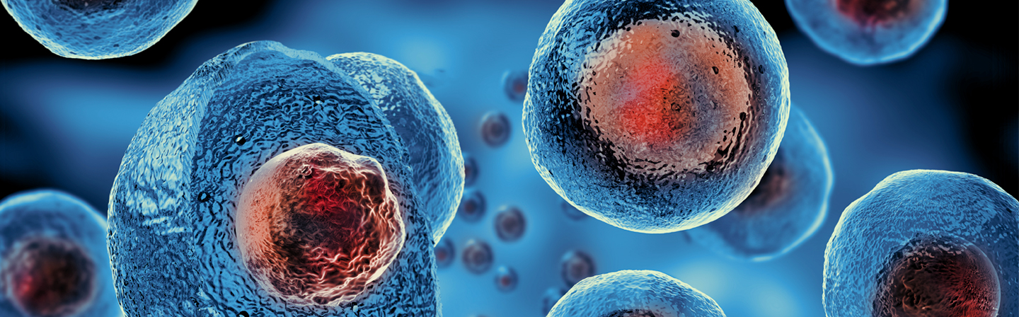
Confocal microscope for rapid detection of tumor boundaries during surgery

Project period: 04/2018-12/2022

© Fraunhofer IPMS
Demonstrator of the microscope at the Fraunhofer Center MEOS in Erfurt, Germany.

Every year, approximately 500,000 people in Germany are diagnosed with cancer. Diagnostics and treatment are developing steadily, but today it is still virtually impossible for doctors to determine the success of a tumor resection in a very short time during surgery. Thus, after surgical removal of a tumor, it is common practice to take a tissue sample from the wound margin and have it pathologically examined in the laboratory to ensure that all tumor cells have been removed. This procedure takes up to 20 minutes. What would be desirable is a method that can be used quickly, reliably and directly on site in the operating room. Therefore, employees of Fraunhofer IPMS and Fraunhofer IZI have jointly developed a new confocal microscope for intraoperative tumor diagnostics at the Fraunhofer Center MEOS.

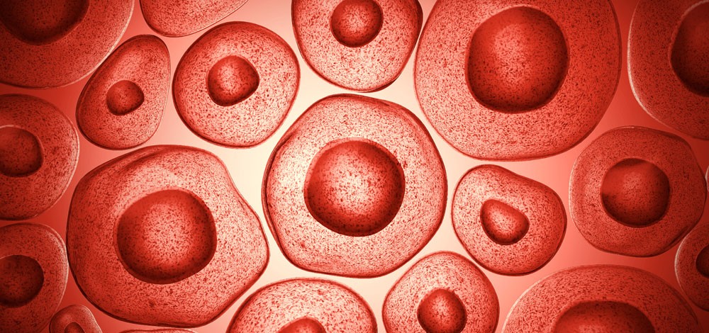
The distinction between healthy tissue and tumor tissue is often not easy to make. The goal of tumor surgery is to create tumor-free resection margins while increasing the preservation of surrounding healthy tissue. This is currently complicated by the fact that current technical solutions do not allow three-dimensional imaging of the tumor margin. Histological analysis during surgery is feasible, but it is time-consuming and not feasible for all tissue types (e.g., bone). Therefore, there is a great need for an easily applicable, optical method that differentiates healthy tissue structures from tumor tissue intraoperatively and within a fast time frame. 

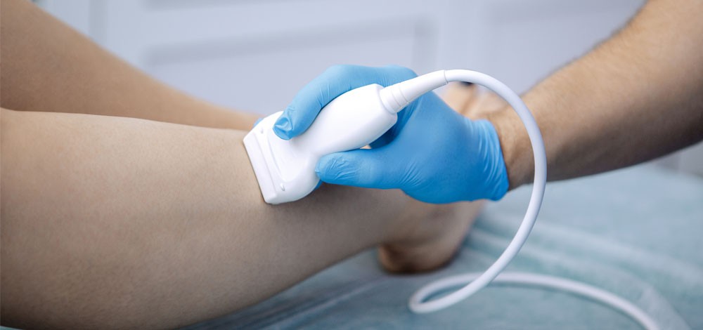
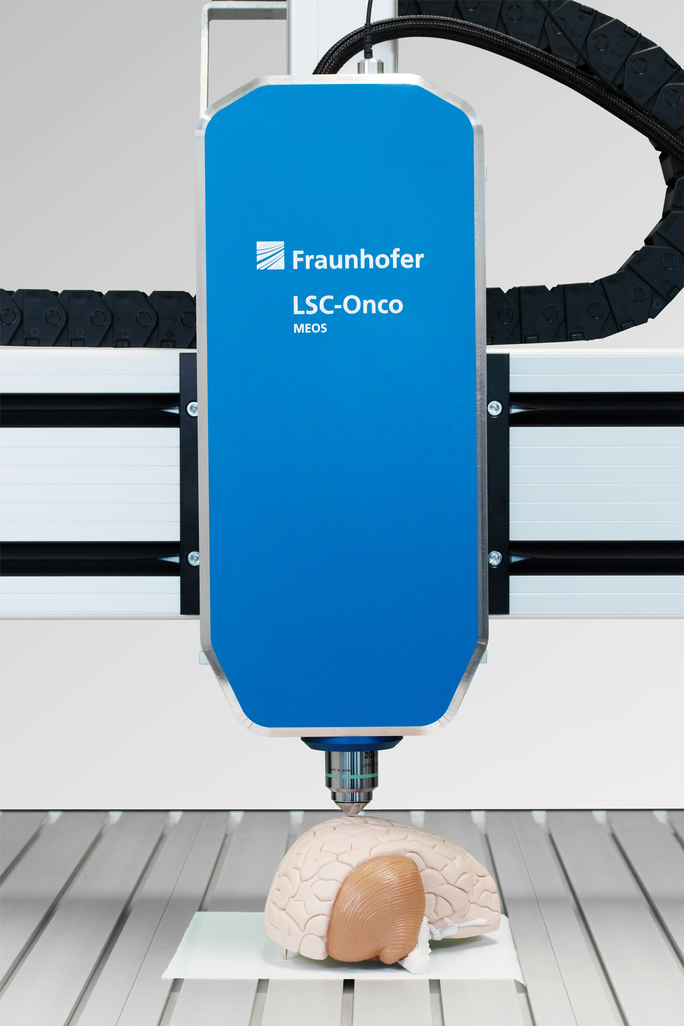
In the Fraunhofer Center MEOS, employees of Fraunhofer IPMS and Fraunhofer IZI have jointly developed a MEMS-based laser scanning microscope and a fluorescence marker method of tumor cells. The aim is to localize tumor boundaries in the best possible way to ensure the complete preservation of e.g. brain cells and arteries during neurosurgical interventions. In the first step, the tumor margin must be stained for this purpose. Here, a special method for the specific staining of tumor cells using fluorescence-labeled antibodies at the cell culture level is used, which was developed by Fraunhofer IZI staff. An image of the cut surface is then taken through the confocal microscope. The core of the microscope is a scanning mirror developed at Fraunhofer IPMS, which allows the light to be deflected in the x- and y-directions, thus generating an image practically in real time. This allows a lateral resolution < 1.0 μm to be achieved in the fluorescence image with a field size of 200 x 200 μm² (960 x 960 pixels). For sectional images, the system is equipped with a z-shifter with a max path length of 2000 μm and 5 nm min step size.

In 2021, a demonstrator of the microscope was set up and successfully tested at the Fraunhofer Center MEOS in Erfurt. For this purpose, tissue samples were provided by the application partner, Helios-Klinikum in Erfurt. Future work will focus on the use of artificial intelligence (AI) for automated detection of tumor resection margins, robotics to create an assistance system for surgical staff, and system adaptations for transfer to a clinical environment.

Ultrasound-based imaging in medical technology and industrial metrology

Applications for bio and health

In medical technology and industrial measurement technology, sonography is an established field of analysis. The use of ultrasonic transducers in the form of ultrasonic arrays is of decisive importance for these imaging techniques. The majority of ultrasound arrays manufactured in medical technology today use piezoelectric ceramic lead zirconate titanate (PZT), exploiting the reverse piezoelectric effect, to generate sound. However, high-frequency, high-resolution or contactless air-coupled arrays based on PZT are expensive and complex to manufacture. In addition, the toxic materials used are only sustainable to a limited extent (RoHS conformity).

Capacitive micromechanically manufactured ultrasonic transducers (CMUT) open up new possibilities here. Micromechanical manufacturing processes enable the economic production of corresponding ultrasound arrays for the first time. Furthermore, the high miniaturisation enables the use of CMUTs in invasive applications (e.g. intravascular ultrasound, IVUS). The results of the developments to date indicate a good property profile for CMUTs for the production of high-frequency arrays. The CMUT devices offer:

  • very high acoustic bandwidth
  • extremely low mechanical coupling between the elements
  • good adaptability to water and air
  • integration together with electronic components (ASIC)
  • no toxic materials (RoHS conformity)

High bandwidth and low coupling between channels are fundamental condition for imaging that can match the standards of conventional ultrasound imaging. Fraunhofer IPMS' highly integrated MEMS technology makes it possible for the first time to connect the signals of an array on site with readout electronics to achieve a simple and compact contacting of the elements. By using the monolithic connection technology between sensor and electronics, highly planar surfaces can be realised as a contact to the medium.

MEMS-based confocal fluorescence microscope

Applications for bio and health

With a confocal fluorescence laser scanning microscope, the sample is irradiated point by point and the fluorescence radiation excited in the sample is measured. In addition to capturing horizontal sectional images, this technology enables the production of 3D models of structured surfaces and fluorescent samples. The main areas of application are in biological and medical research as well as industrial quality assurance. However, due to the areas of application in research, these are usually complex stationary and correspondingly cost-intensive instruments.

Fraunhofer IPMS has therefore developed a robust and portable MEMS-based fluorescence laser scanning microscope using standard optics. This is made possible by integrating a 2D microscanning mirror developed in-house.

Structured illumination for microscopy - targeted illumination of biomedical samples to reduce light-induced damage

Applications for bio and health

Privacy warning

With the click on the play button an external video from www.youtube.com is loaded and started. Your data is possible transferred and stored to third party. Do not start the video if you disagree. Find more about the youtube privacy statement under the following link: https://policies.google.com/privacy

The modern method of light microscopy faces a major challenge: the permanent illumination of the entire sample with relatively high brightness causes damage from light radiation. To reduce this phototoxic effect and protect the sample, the illumination must be selective and limited to the area to be examined.

Fraunhofer IPMS is developing an innovative solution to this problem as part of the Fraunhofer Project Center MEOS: optical modules for high-resolution microscopy that can selectively control light. This is made possible by the patented micromirror arrays of Fraunhofer IPMS.

Up to several million mirrors are located on a semiconductor chip and these miniature deflection mirrors can modulate and direct the light. A specially developed optical setup using two micromirror arrays enables control of both the illumination pattern and the angle of the light incident on the sample. In this way, biological samples can be examined as part of a very careful, high-resolution process.

Mobile analysis systems

Applications for bio and health

Improved imaging in point-of-care diagnostics requires very compact systems with high optical magnification, which are realised by combining multi-aperture optics with novel MEMS drive principles. Systems with optical resolution in the sub-μm range are possible, which are fabricated by integration at wafer level. This allows robust diagnostics while reducing the time required for a single analysis. The basic optical imaging principle requires relative movement of the multi-aperture optics to the object for magnified image acquisition.

A first fully functional demonstrator still uses piezo components for this purpose. In order to further miniaturise the system, these will be replaced by special MEMS actuators, so-called inchworm drives, for which a patent application has now been filed and which use the NED drive principle developed at Fraunhofer IPMS. The integration of these MEMS actuators into the demonstrator will take place in the near future.