 Fraunhofer Institute for Photonic Microsystems
MEMS mirrors for genetic research
Researchers at the Fraunhofer-Institut für Photonische Mikrosysteme IPMS (Fraunhofer Institute for Photonic Microsystems) in Dresden have developed a programmable microelectromechanical (MEMS) chip which can divert light of varying wavelengths at ultra-high speed and with micrometer-accuracy. Installed in an optical microscope, this technology can be used to illuminate multiple targeted areas, which can be smaller than single cells and thereby stimulate specific light sensitive molecules as ensemble . In addition it is possible, by using a second chip, not only to select specific areas precisely but also the angle at which these are illuminated. This technique is able to reach objects that appear as structures to be highlighted with even greater precision and to significantly reduce the many undesired environmental influences.
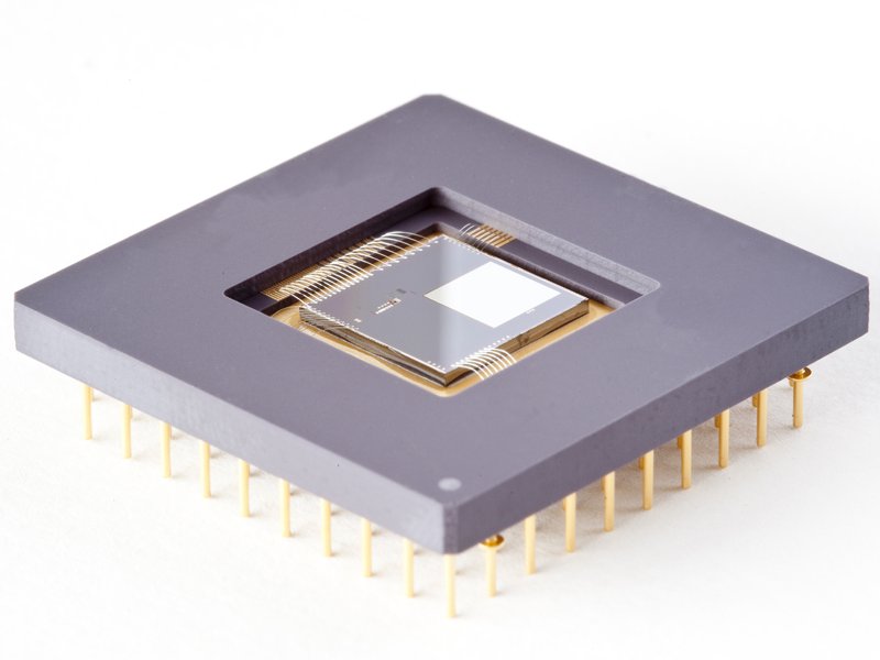
A single MEMS chip consists of an array of 65,536 separate micro mirrors which can each be tilted separately and virtually in a continuous way . By controlling the deflection of all mirrors it is possible to distribute the angle of incidence and the intensity of the light with up to 1,000 changes per second over the entire matrix area. In order to make the benefits of this MEMS technology amenable for optical microscopes, researchers at Fraunhofer IPMS team up with the manufacturer of optical systems, IN-VISION Digital Imaging Optics GmbH in Austria, and scientists of the plateforme d'imagerie dynamique (PFID) at the Institut Pasteur, in France. The aim of the French consortium’s leader is to apply this new combination of optics and genetics to influence the expression of individual genes in cells or organs of zebrafish embryos and fruit fly larvae. Such an intervention will make it possible to study the influence of specific genes on the development of organisms with far greater precision than before. The system is also intended to be used to activate neurons by means of genetically modified, light sensitive ion channels and thereby explore the function of individual neural networks in cerebral tissue.
This research collaboration between Fraunhofer and the Institut Pasteur is being supported by the German Federal Ministry for Education and Research and the French National Research Agency within the framework of the Inter Carnot Fraunhofer Programme. A first experience of the system and the latest research results will now be presented to the public on the joint exhibition stand no. 224 at the specialist conference and SPIE Photonics Europe in Brussels from April 14 to 17, 2014.
About Institut Pasteur, Imagopole (PFID)
The PFID (plateforme d'imagerie dynamique) is a research technology platform (core facility) providing expertise and support in optical imaging methods for researchers at the Institut Pasteur. Our activities include service rendering, training, technology-driven research and technology development. These activities are highly multi-disciplined, and collaborative, with the mission goal focused on the use of quantitative imaging and analysis to understand the processes of cell/tissue-biology, and their usurpation by infection and disease.
The PFID's R&D activities are founded upon the need to develop optical imaging methods that bring new understanding of host-pathogen interactions at the tissue, cellular and molecular levels. In this context, we aim to satisfy the continuous need for novel techniques allowing probing infection, and extrapolating quantitative information on spatiotemporal dynamics in situ. On the other hand we are also interested to push the limits of existing approaches aiming to enhance their performance thereby broadening their experimental utility. The PFID is especially interested in the development of in situ high-content imaging techniques and their application to infection, cell biology, cellular microbiology, and microbiology.